Related pages
In this section
-
Key findings
-
Source localization
-
Imaging of brainstem and cerebellum
-
Early visual processing
-
Visual perception
-
Face processing
-
Attention
-
Music perception
-
Sleep
-
Eye movements
-
Somatosensation
-
Single trial variability
-
Patients
-
Data analysis overview
Visual perception
To see the detailed figure legends hover the mouse over the figures
Effects of context on the visual processing
The visual system rapidly completes a partially occluded figure. We probed this completion process by using a "same-different" priming task where test figures are preceded by a sequence of two figures, a prime or control figure followed by an occluded figure.

Priming effect. Stimuli, and the localization and time course of the right fusiform gyrus
(FG) activity. Time courses were averaged for each of the 10 first stimulus types (shown in different
colours) for all 10 subjects. Simple figures led to significantly reduced activity in the right FG compared
with control figures between 120 and 200 ms, as highlighted by the gray box.
Priming reduced significantly the activation levels in the right fusiform gyrus between 120 and 200 ms after stimulus onset (gray box). This modulation of activity may be the neural correlate of priming and suggests this area as a hub for different occluded image interpretations in the early stages of perceptual processing.
L. C. Liu et al., Neuroscience 141, 1585-1597 (2006). PDF >>
Neural processing of illusory figures
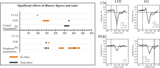
"Kanizsa" illusory figures were presented to subjects at central and peripheral locations of the visual field in three different task conditions: a classification response task, in which subjects responded to illusory figures, but not to control figures; an onset response task in which subjects responded whenever a stimulus was shown; and a no response task, where subjects viewed the stimuli passively.

Processing of illusory figures. (On the left) The latencies of significant, as determined
by ANOVA, effects of illusory figures and tasks in areas V1/V2, lateral occipital cortex (LOC) and fusiform
gyrus (FG), and (on the right) the activation time courses of LOC and FG. The results are shown separately
for central (upper rows, CM) and peripheral (lower rows, PERI) visual field presentations.
Completion of the illusory figures at the fovea and periphery is accomplished using different brain mechanisms. Completion at the fovea is realized in a more automatic manner at the early stages of visual information processing, while at the periphery it takes place at the later stages and somewhat requires involvement of attentional processes. In addition, the illusory figure processing at the fovea engages more the lateral occipital cortex (LOC), whereas at the periphery the fusiform gyrus (FG) is mostly involved.
A. A. Bakar et al., Human Brain Mapping 29, 1313-1326 (2008). PDF >>