Related pages
In this section
-
Key findings
-
Source localization
-
Imaging of brainstem and cerebellum
-
Early visual processing
-
Visual perception
-
Face processing
-
Attention
-
Music perception
-
Sleep
-
Eye movements
-
Somatosensation
-
Single trial variability
-
Patients
-
Data analysis overview

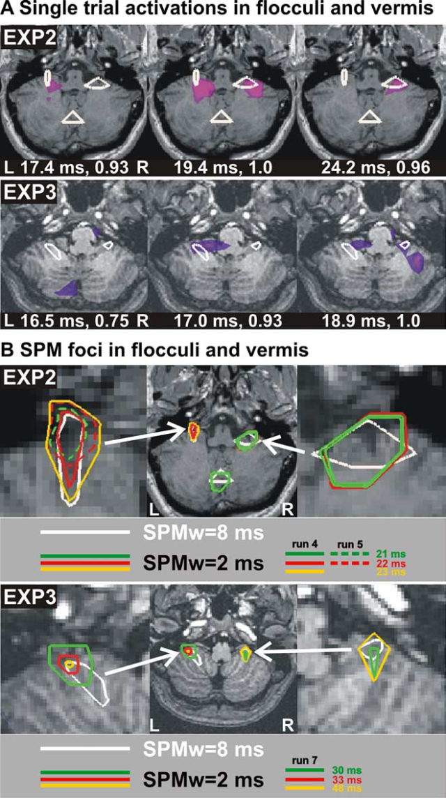
Cerebellum spike activity and statistically significant foci shown on axial MRI cuts at the level
of the flocculi and the dorsal vermis. (A) The top half shows peaks of activity from a single trial
of saccade from center to left at 17.4, 19.4, and 24.2 ms. The MFT source analysis result shown in
pink/purple is normalized in strength to 1 (middle panel). White contours are early statistically significant
foci computed using an 8 ms running window and the pre-saccadic period (-190 to -160 ms) as baseline. The
bottom half shows the same for a trial for saccade to the left at 16.5, 17, and 18.9 ms. Note that the
single-trial activations show rapid interplay between the flocculi whose positions are in good agreement with
both the anatomical definition and the statistical results. (B) Statistically significant foci for saccades
from center to left. Top row: results from experiment 2 with statistical comparisons performed with 2 ms
running window and the pre-saccadic period (-190 to -160 ms) as baseline. For reference, white contours in
(A) are included. Green centered at 21 ms, red at 22 ms, yellow at 23 ms, all showing precise localization at
the flocculi and vermis. Solid color contours are results from run 4, and dotted ones are from run 5, showing
rapid habituation of vermis and right flocculi leaving only the ipsilateral (left) floculus focus. Bottom
row: the same arrangement for experiment 3, bilateral flocculi are seen active at 30 ms, at 33 ms only in the
left flocculus, and at 48 ms in both flocculi again. L, Left; R, right.
The figure shows the rapid transition of instantaneous activations (MEG spikes) between flocculi and vermis (figure A), and the first statistically significant (P < 0.00001) loci in these two areas (figure B), shortly after saccade onset. These statistically significant changes in activity are consistent with the roles of these cerebellum structures in controlling initial eye position and monitoring of saccade progress. Each flocculus was more active for ipsilateral saccades, but overall MEG spikes were identified in both flocculi for saccades to the left and right. The vermis activation was restricted to the dorsal vermis, uvula, and the nodulus.
Multiscale bistable intentionality
The current density vectors, i.e. estimated neural sources contributing to the MEG signal are essentially confined in two dimensions, and therefore their variation can be conveniently quantified and displayed using circular statistics (N. I. Fisher (1993) Statistical analysis of circular data. Cambridge, MA: Cambridge UP.)
The next two figures show results from experimental conditions where two opposite saccade directions are possible before the GO signal. The direction of the saccade is provided together with the GO signal, so the subject has to be prepared to make a saccade in any of the two directions.

Spike activity in frontal eye fields (FEF) and a sharpening of current density directions just
before or around saccade onset. (A) Instantaneous single-trial activations from a saccade from
center to right, with FEF region of interest marked by white circles. Spikes from both FEFs are seen. Only on
one occasion a spike is captured in two successive time slices (separated by 0.48 ms). Top row: experiment 2,
bottom row: experiment 3. (B) Circular statistics for saccades to the left (left panel) and right (right
panel). The white arrow is the significant direction for the vectors as defined by circular statistics.
Multiple arrows signify multiple directions. Top: computed from two pre-saccadic periods (-200 to -100 ms and
-90 to -10 ms) in experiment 2. In each FEF, a bimodal distribution is seen, except for the last 90 ms before
the saccade, when the distribution is nearly unimodal for the FEF contralateral to the saccade direction of
the saccade (marked by yellow stars). Bottom: computed from around two saccade onset periods (-40 to 0 ms and
0-40 ms) in experiment. A pattern of activity similar to that for experiment 2 is found, especially for
saccades to the right.
Figure A shows the instantaneous MFT single trial activity (current density vectors) for frontal eye fields (FEF). Note rapid interchanges between left and right FEFs. When two saccade directions are possible as in the above example, regional activations are associated with bimodal current density distributions over long periods, thus keeping both options alive, a state we call multiscale bistable intentionality. Examples using, circular statistics, of such a state are shown for activity in the contralateral FEF (figure B above this paragraph) and ipsilateral brainstem gaze centers and flocculi (figure below). Multiscale bistable intentionality requires two simultaneous sustained directional signals corresponding to each of the two possible options.

Multiscale bistable intentionality in the gaze centers and flocculi. Circular statistics
for experiments 2 and 3 at three periods, -60 to -20 ms, -20 to 20 ms, and 20-60 ms relative to saccade
onset. A?D, experiment 2 (A, B) and experiment 3 (C,D), left (L) and right (R) gaze center. The top row shows
diagrams from saccades from center to left; the bottom row shows diagrams from saccades from center to right.
White numbers below each diagram are the largest vector for that picture normalized to the strongest vector
in the whole figure. Experiment 2 (E, F ) and 3 (G, H) left and right flocculi for ipsilateral saccades. For
reference, the regions of interest definitions for the gaze centers and flocculi are also shown on the left
and right MRIs, respectively. Note the similarity of pattern across subjects with activity for the time
during saccade, becoming focal and unidirectional, with different vector directions for different saccades.
Also note the increasing current density as saccade occurs. I-L, Circular statistics diagrams computed for a
control area (auditory cortex) in the same hemisphere and periods as the flocculi.
The figure below shows results from experimental condition where the saccade direction is provided to the subject long before the GO signal. So the subject has to be prepared for a saccade in only one direction.

The frontal eye field activity reflects the task demand. Circular statistics for frontal
eye fields from experiment 1, GO and NOGO trials after the cue heralding the direction of saccade. The top
half (in green boxes) is for GO trials, and the bottom half (in red boxes) is for NOGO trials, and this
information is provided 1-3 s before the cue. The actual saccade for the GO trials is initiated 1-3 s after
the cue. In each group of pictures, the top row shows the circular statistics for 100-400 ms after the onset
of the cue, and the bottom row shows the circular statistics over a narrower sub-range 100 ms long. The
yellow stars show the contralateral frontal eye field (FEF) relative to the saccade direction. The direction
of the current density vector becomes more unimodal for contralateral FEF in GO trials but remains random for
ipsilateral FEF in GO trials and the NOGO condition.
When only a single saccade direction is possible as in the example above, in GO trials the directional information about a future saccade quickly transforms a random distribution of current density into a unidirectional distribution well before any saccade is initiated.
Summary
Preparation for impending saccade begins as soon as relevant information becomes available, in some cases several seconds before the saccade. Cues providing partial information initiate competing motor programs for as yet undecided future actions that are maintained until cues with new information resolve the uncertainty.
A. A. Ioannides et al., Journal of Neuroscience 25, 7950-7967 (2005). PDF >>
Random eye movements (REM) in sleep
We recorded the MEG signal from three subjects before, during and after eye movements cued to a tone, self-paced, awake and during rapid eye movement (REM) sleep.

Across-subject statistical parametric maps (SPM) for leftward eye movements. The loci of
common changes are identified after the data from each subject are transformed into the Talairach space. The
common loci for the three subjects are displayed after back-transforming the results and projecting them onto
the MRI of one subject (red - increase, blue - decrease; the time interval and the threshold P value are
printed inside each figurine). Axial and sagittal MRI slices for three successive SPMs leading to saccade
onset (the contrast is REM sleep versus eyes closed awake state using 12 ms running windows). The SPMs show a
sequence of relative increase of right hemisphere activity in REM beginning in the orbitofrontal cortex and
amygdala (A), followed by activity in the parahippocampal gyrus (B) and finally pontine nucleus (C).
An orbitofrontal-amygdalo-parahippocampal-pontine neural activity sequence, possibly related to emotional activation during REM sleep, was identified in the last 100 ms leading to the REM saccade, but not linked to saccade initiation.
A. A. Ioannides et al., Cereb. Cortex 14, 56-72 (2004). PDF >>